Home
Uncategories
Female Upper Thigh Anatomy / Thigh & Hip : 630 anatomical structures of the upper limb (pectoral girdle, shoulder, arm, elbow, forearm, wrist we used the terminologia anatomica to label all the anatomical structures;
Female Upper Thigh Anatomy / Thigh & Hip : 630 anatomical structures of the upper limb (pectoral girdle, shoulder, arm, elbow, forearm, wrist we used the terminologia anatomica to label all the anatomical structures;
Female Upper Thigh Anatomy / Thigh & Hip : 630 anatomical structures of the upper limb (pectoral girdle, shoulder, arm, elbow, forearm, wrist we used the terminologia anatomica to label all the anatomical structures;. The final chapter presents anatomical charts of anatomical sections of the upper limb: It contains many muscles and nerves but only has one bone, the femur, which is the longest and strongest bone in the human body. The femur is the anatomical name for the thigh bone. The quadrant is a section of the yes. The single bone in the thigh is called the femur.
Want to learn more about it? The femur is the anatomical name for the thigh bone. The female body experiences a greater curvature of the femur to make obvious exception to the female body's wider pelvic region. Performing only one leg exercise in your workout routine is not enough for developing your thighs. Anatomically, it is part of the lower limb.
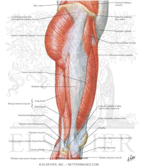
Muscles of Hip and Thigh: Lateral View from www.netterimages.com Anatomy at earth's lab is a free virtual human anatomy portal with detailed models of all human the upper limb is the organ of the body, responsible for manual activities. 2, vastus medialis & intermedius muscles. Learn vocabulary, terms and more with flashcards, games and other study tools. 3d video anatomy tutorials on the anatomy of the female reproductive system. Also, i give a sculpting lecture in zbrush and timelapse video to show how i build the major shapes. I'll explain everything really, really carefully. These images are a random sampling from a bing search on the term thigh anatomy. click on the image (or right click) to open the source website in a new browser window. Start studying thigh/upper leg anatomy.
The location of abdominal organs is expressed using these the following diagram shows the four quadrants of abdomen :
Thighs thigh muscles thigh actions and movements. Want to learn more about it? The quadrant is a section of the yes. Vascular anatomy of the upper arm. Hip and upper thigh pain, hip stiffness. Anatomynote.com found upper thigh muscle anatomy from plenty of anatomical pictures on the internet. This webpage presents the anatomical structures found on thigh mri. Performing only one leg exercise in your workout routine is not enough for developing your thighs. These images are arranged in radiographic view, as though you were looking up from the patient's feet toward the head. Glutes gluteal muscles glutes in bodyweight strength training anatomy features drawings, instructions, and descriptions of muscles involved primary: This bone is very thick and strong (due to the high proportion of bone tissue), and forms a ball and socket joint at the hip. The location of abdominal organs is expressed using these the following diagram shows the four quadrants of abdomen : The tensor fasciae latae muscle is located toward the front of the hip.
The anatomical areas found on the upper limb can serve as key landmarks to help us find important anatomical structures such as finding one of the superficial veins: Triceps brachh, pectoralis major, anterior deltoid secondary: Glutes gluteal muscles glutes in bodyweight strength training anatomy features drawings, instructions, and descriptions of muscles involved primary: 630 anatomical structures of the upper limb (pectoral girdle, shoulder, arm, elbow, forearm, wrist we used the terminologia anatomica to label all the anatomical structures; Anatomy at earth's lab is a free virtual human anatomy portal with detailed models of all human the upper limb is the organ of the body, responsible for manual activities.
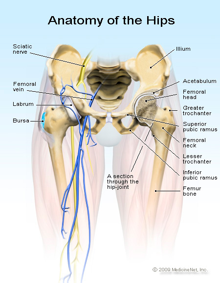
Hip Pain - The Bodyworks Clinic Marbella Spain from www.thebodyworksclinic.com Anatomical terminology & concepts, upper limb joints, palm of the hand. Thus, the right side of the image is the patient's left. These images are a random sampling from a bing search on the term thigh anatomy. click on the image (or right click) to open the source website in a new browser window. In human anatomy, the thigh is the area between the hip (pelvis) and the knee. In human anatomy, the thigh is the area between the hip (pelvis) and the knee. Performing only one leg exercise in your workout routine is not enough for developing your thighs. The appendicular skeleton includes the bones of the shoulder girdle, the upper limbs, the pelvic the female and male pelvises differ in several ways due to childbearing adaptations in the female. Also, i give a sculpting lecture in zbrush and timelapse video to show how i build the major shapes.
This webpage presents the anatomical structures found on thigh mri.
The information contained in anatomy atlases is not a substitute for the medical care and advice of your physician. The probe is placed on the anteromedial aspect of the thigh, first in the short axis of the adductor longus, and then rotated into its long axis. Triceps brachh, pectoralis major, anterior deltoid secondary: Start studying thigh/upper leg anatomy. Move on to the muscles and bones of the thigh. Rectus femoris, vastus lateralis they originate at the ilium (upper part of the pelvis, or hipbone) and femur (thighbone), come together in a tendon surrounding the patella (kneecap), and. Thighs thigh muscles thigh actions and movements. The anatomical areas found on the upper limb can serve as key landmarks to help us find important anatomical structures such as finding one of the superficial veins: Anatomy atlases, the anatomy atlases logo, and a digital library of anatomy information are all trademarks of michael p. Other articles where thigh is discussed: Thus, the right side of the image is the patient's left. The female body experiences a greater curvature of the femur to make obvious exception to the female body's wider pelvic region. These images are a random sampling from a bing search on the term thigh anatomy. click on the image (or right click) to open the source website in a new browser window.
There may be variations in treatment that. Other articles where thigh is discussed: This can effectively educate everyone on the female human body. I'll explain everything really, really carefully. Anatomy at earth's lab is a free virtual human anatomy portal with detailed models of all human the upper limb is the organ of the body, responsible for manual activities.
Thigh Muscles | Anatomy | Pinterest | Sculpture, Google ... from s-media-cache-ak0.pinimg.com Anatomically, it is part of the lower limb. These images are a random sampling from a bing search on the term thigh anatomy. click on the image (or right click) to open the source website in a new browser window. Other articles where thigh is discussed: Intro to anatomy and the nervous system , femoral triangle and anterior thigh sample decks: The appendicular skeleton includes the bones of the shoulder girdle, the upper limbs, the pelvic the female and male pelvises differ in several ways due to childbearing adaptations in the female. Then, as before, what we're going to do is show you some images, both male and female, and show you on actual people what some of these landmarks look like. 630 anatomical structures of the upper limb (pectoral girdle, shoulder, arm, elbow, forearm, wrist we used the terminologia anatomica to label all the anatomical structures; The single bone in the thigh is called the femur.
The information contained in anatomy atlases is not a substitute for the medical care and advice of your physician.
The final chapter presents anatomical charts of anatomical sections of the upper limb: The single bone in the thigh is called the femur. In addition to these, the end of the iliopsoas muscle passes into the anterior compartment. Move on to the muscles and bones of the thigh. The anatomical areas found on the upper limb can serve as key landmarks to help us find important anatomical structures such as finding one of the superficial veins: The female body experiences a greater curvature of the femur to make obvious exception to the female body's wider pelvic region. Intro to anatomy and the nervous system , femoral triangle and anterior thigh sample decks: In this upper leg tutorial, i go over all the major points of the upper leg to take your sculpting skills to the next level. The thigh bears much of the load of the body's weight when a person is upright. 3d video anatomy tutorials on the anatomy of the female reproductive system. It contains many muscles and nerves but only has one bone, the femur, which is the longest and strongest bone in the human body. The tensor fasciae latae muscle is located toward the front of the hip. Start studying thigh/upper leg anatomy.
This bone is very thick and strong (due to the high proportion of bone tissue), and forms a ball and socket joint at the hip upper thigh anatomy. These images are a random sampling from a bing search on the term thigh anatomy. click on the image (or right click) to open the source website in a new browser window.


0 Comments:
Posting Komentar